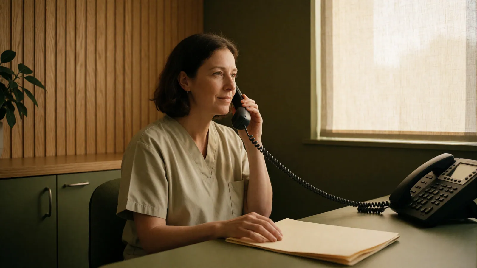
Some things should not wait for the next free routine appointment. Some things should not wait for the next free routine appointment. Telephone the practice and say what is happening, or go to an emergency room, if you have a mole or a mark that has changed shape, colour or size, that has become raised or uneven, or that bleeds, itches or will not heal; a sore anywhere that has not healed in three weeks; a rash that is spreading quickly, especially with fever, blistering, or peeling skin; a painful rash in a band on one side of the body, particularly near an eye; redness that is hot, swollen, tender and spreading, which can be a deep skin infection; a new rash after starting a medicine, especially with mouth or eye involvement; or a child under three months with any widespread rash. If you have been treated here and something is worse rather than better after a procedure, telephone us rather than waiting for the review appointment. We would far rather field a call that turns out to be nothing.
Melanoma arises from the pigment-producing cells in skin. It is far less common than the basal and squamous cell cancers, and it is the one that can travel elsewhere in the body, which is the entire reason it is treated differently. About half of melanomas appear as a new mark rather than in an existing mole, which is why the instruction to watch your moles, on its own, is incomplete advice — the thing to watch for is anything new or changing, mole or not.
The number that governs everything afterwards is how deep it has grown, measured in millimetres by a pathologist. That is why a suspicious pigmented lesion is removed whole with a margin rather than shaved or partially sampled: a partial sample can miss the deepest part and mislead everybody about how serious it is. It is also why time matters. Depth accumulates with time, and the difference between a melanoma found this month and the same one found next year is frequently the difference between an outpatient excision and a course of treatment.
What this practice does is the recognising and the biopsy. If the report comes back as a melanoma, your care moves to a cancer centre, because staging, wider surgery, lymph node assessment and any further treatment need a team rather than a dermatologist. We make that referral ourselves and we stay involved for the skin surveillance afterwards, which is lifelong.
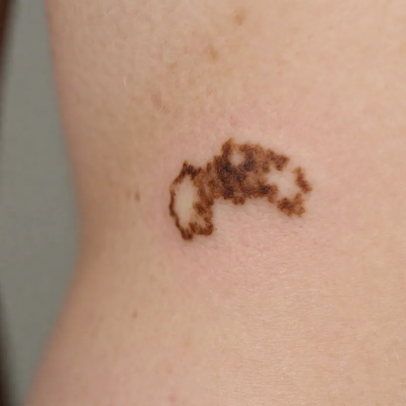
The changes that should bring you in
This is a list of reasons to book an appointment. It is not a list you can use to rule anything out.
A new pigmented mark after about thirty
New moles are ordinary in childhood and adolescence and become less ordinary with age. A genuinely new brown or black mark in an adult is worth looking at, particularly if it is growing.
Any mole that is changing
Getting larger, getting darker, developing a second or third colour, becoming raised where it was flat, developing a ragged or blurred outline, or becoming itchy, crusted or bleeding. Change in any direction, over weeks to months, is the signal.
The one that does not match your others
This is the most practical instinct for a patient to use, and it works better than measuring. Your moles broadly resemble each other; the outlier is the one to have looked at.
A firm growing lump, even a symmetrical, evenly coloured or pink one
The fast-growing nodular form frequently breaks every rule of the familiar checklist — it can be small, round, one colour, and not brown at all. What it does is grow, over weeks rather than years, and feel firm. Do not talk yourself out of an appointment because it looks tidy.
A dark band under a fingernail or toenail
Particularly a single nail, particularly if the band is widening, and especially if the pigment extends onto the skin at the base of the nail. This form is the one most often diagnosed late, and it is disproportionately a risk on darker skin.
A dark patch on a palm, a sole, or inside the mouth
Sites that are frequently skipped in a rushed examination and that matter most for patients with deeply pigmented skin, where melanoma is uncommon but is more likely to occur in exactly these places.
How it is diagnosed here
Nothing on this page can diagnose you. This is what the actual process looks like.
Examination and dermoscopy first
Which raises or lowers suspicion but does not settle it. A dermatoscope is very good at telling an ordinary mole from an unusual one; only a pathologist can say what an unusual one actually is.
Excisional biopsy, whole, with a narrow margin
Under local anaesthetic, taking the entire lesion and a small rim of normal skin around and beneath it, closed with sutures. This is the correct technique because the pathologist needs the deepest part. It is not the same as a shave, and where somebody has had a suspicious mole shaved elsewhere the depth is often unknowable afterwards.
The report takes about a week
Sometimes longer, if the pathologist orders additional stains — which is thoroughness rather than bad news. We telephone every result, and you are told before you leave how and when that call will come.
If it is a melanoma, the referral is made for you
To a cancer centre, with the pathology report and the images. That team decides on wider excision, whether the lymph nodes need assessing, and any further treatment. We do not manage that here and we will not pretend otherwise — see /scope-of-care.html.
Skin surveillance afterwards continues here, for life
Having had one melanoma is the strongest single predictor of having another. Follow-up examinations are more frequent, more thorough, and include the scar, the surrounding skin and the lymph node areas. This part we do.
What raises the risk
Risk factors change how closely somebody should be watched. Not one of them means you will develop this.
Ultraviolet exposure, particularly burning
Episodes of blistering sunburn, especially in childhood, and the use of sunbeds. Intermittent intense exposure appears to matter more for melanoma than steady outdoor work, which is a different pattern from the other skin cancers.
Fair skin that burns rather than tans
Along with red or blond hair, freckling and light eyes. This is the classic risk group and the one most likely to be examined regularly already.
Many moles, or unusual-looking moles
A large number of ordinary moles, or a smaller number of atypical ones, both raise risk and both are reasons for a mapped baseline examination.
Family history in a close relative
A parent, sibling or child with melanoma raises your risk meaningfully and lowers the threshold for regular examination. Tell us — it changes the interval we recommend.
A previous skin cancer of any kind
And a suppressed immune system, whether from transplant medication or long-term immunosuppressive treatment for another condition.
Darker skin does not exclude it
It is much less common, it occurs in different places — palms, soles, nail beds — and it is diagnosed later, which is what worsens outcomes. Every skin examination here includes those sites on everybody.
Melanoma in western Queens
The least common of the major skin cancers and the one that matters most, because it can spread. Found early it is usually dealt with by removing it. This page is about recognising change and getting seen — not about self-diagnosis.
Every appointment here starts the same way: a proper history, an examination in good light, and a named diagnosis said out loud and written down for you to take home. If there is genuine doubt you will be told that, and told what would settle it — usually a biopsy. You leave with a plan that has a strength, a site, a frequency and a date to stop or come back on it, and, where the condition is one that relapses, what to do on the first day of the next flare. If the honest answer is that this is better left alone, or that it belongs somewhere other than here, that is what you get instead, and it costs exactly the same.

This page is not medical advice, and it is not a diagnosis. Nothing on this website is medical advice for you in particular, and it is not a diagnosis. These pages describe conditions in general terms so that you arrive knowing what the questions are — they cannot tell you which condition you have, because that requires somebody to look at your skin, feel it, ask you a hundred questions and frequently take a sample of it. Two conditions on this website can look identical in a photograph and need opposite treatments. If something on your skin is new, changing, painful or not healing, the useful next step is an appointment rather than more reading.
No before-and-after photographs, and no promised outcome. There are no before-and-after photographs on this website and there never will be. Every set you have seen somewhere else was chosen from a much larger pile, and the second picture is usually taken in different light, at a different angle, at a different point in a flare cycle. In dermatology the dishonesty is sharper than in most fields, because nearly every condition here fluctuates on its own — photograph any relapsing skin disease at its worst and again at its best and you have a persuasive pair of images that demonstrates nothing about the treatment in between. What we publish instead is what a treatment typically achieves, in how long, in what proportion of people, and how often it does not work.
The risks, plainly. Every treatment described here has side effects, and some carry real risks. Topical steroids thin skin if they are used at the wrong strength on the wrong site for too long, which is why they are prescribed with a site, a strength and a stopping point rather than handed over indefinitely. Light-based and laser treatment can burn, blister and cause pigment change that lasts months, and that risk is higher on deeper skin tones and on recently tanned skin. Any procedure that breaks the skin can bleed, become infected and leave a scar; on the chest, the shoulders and the jawline, scars are more likely to thicken. Several of the medicines used in this speciality require blood tests, and some cannot be taken during pregnancy. We tell you your own risk rather than the average one, and where the honest answer is that a condition is better left alone, that is the advice you will get.
Nothing is diagnosed from a photograph. We do not diagnose skin from photographs, and we do not answer "what is this?" by email, by message or through a form. This is not caution for its own sake. A photograph has no history attached to it, cannot be felt, cannot be stretched or pressed to see whether the colour blanches, cannot be examined under a dermatoscope, and misreports colour badly — which matters enormously on skin that is not pale. The examination that actually distinguishes a harmless growth from an early cancer involves magnification, cross-polarised light and, when there is any doubt at all, a biopsy. An application that offers to tell you whether a mole is dangerous from a picture taken on a telephone is selling reassurance, and reassurance is the one product in this field that is dangerous when it is wrong. Bring the mark to a room where somebody can look at it.
Questions
Melanoma — the questions we are asked most
Can I tell from a picture on the internet whether my mole is a melanoma?
No, and comparing your mark against images online is a genuinely risky habit, because it mostly produces false reassurance. Melanomas vary enormously; several forms look unremarkable, and the fast-growing nodular type frequently looks tidier than the harmless mole beside it. The purpose of this page is to get you to an appointment, not to help you decide you do not need one.
How quickly can I be seen if something has changed?
Telephone and say the word changed. That is triaged differently from a routine appointment and it is not put into the next free routine slot. Do not book a general check-up eleven weeks out and sit on it.
Why remove the whole mole instead of taking a small piece?
Because the measurement that determines everything afterwards is how deep the lesion goes, and a partial sample may not include the deepest part. A shave biopsy of a melanoma can transect it and leave the depth unknowable, which affects the staging and therefore the treatment. Taking it whole avoids that.
Will I need my lymph nodes checked?
That decision belongs to the cancer centre team and depends chiefly on the depth and other features in the pathology report. It is not a decision made here, and anybody telling you the answer before the report exists is guessing.
Does melanoma always look dark?
No. A proportion have little or no pigment and appear pink, red or skin-coloured, which is a common reason for delay — they get mistaken for a spot or a scratch that will not settle. Growth and firmness are the features that matter in those, not colour.
I had a mole removed years ago and the scar has pigment in it. Is that a problem?
It should be examined. Pigment in an old excision scar is sometimes ordinary and is sometimes recurrence, and telling the difference needs dermoscopy and the original pathology report — bring it if you have it.
Related
Often seen alongside this
Skin Cancer & Lesions
Skin Cancer Screening
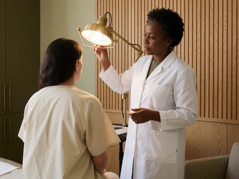
A full-body skin examination in good light with a dermatoscope, taking about forty-five minutes. It finds most of what is present on the day it is done — not all of it — and the part that matters as much as the appointment is what you do between appointments.
Read more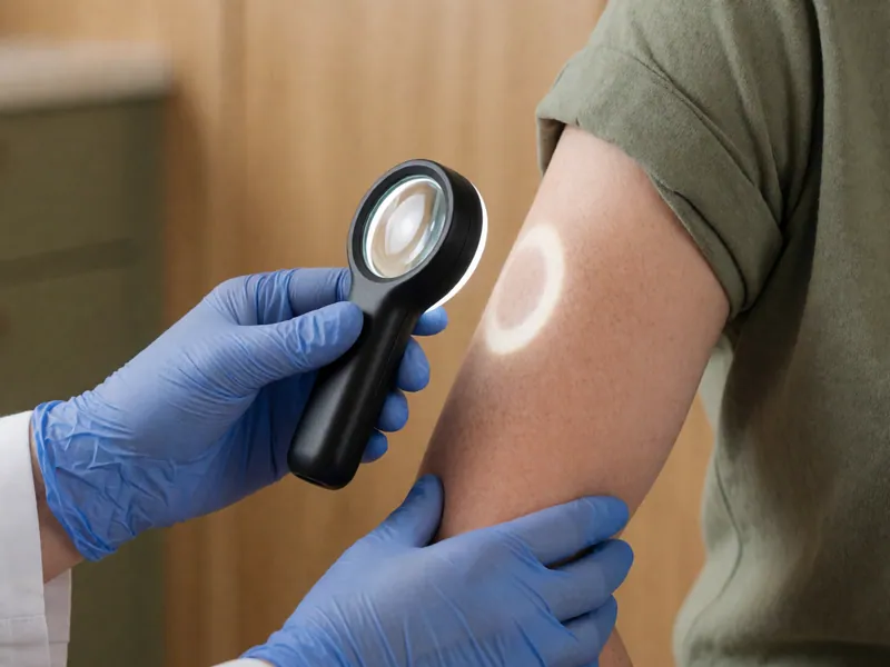
Skin Cancer & Lesions
Mole & Lesion Evaluation
One mark, examined properly: history, naked eye, then dermoscopy. Most moles are entirely ordinary and are left alone with reasons given. Anything genuinely uncertain is sampled rather than watched for a year.
Read more
Skin Cancer & Lesions
Basal & Squamous Cell Cancer
The two common skin cancers. Both are usually slow, both are usually cured by removing them, and both are routinely ignored for a year because they look like a sore that will not heal. Most are treated in this building.
Read moreA named diagnosis and a written plan you leave holding
Book an appointment
Pick a physician and a time from the calendar — it shows only the hours each of them genuinely has free. A new patient appointment is thirty minutes, a full skin examination forty-five, and you leave with a named diagnosis written down. If something on your skin has changed, telephone instead and say that word: it is triaged differently.