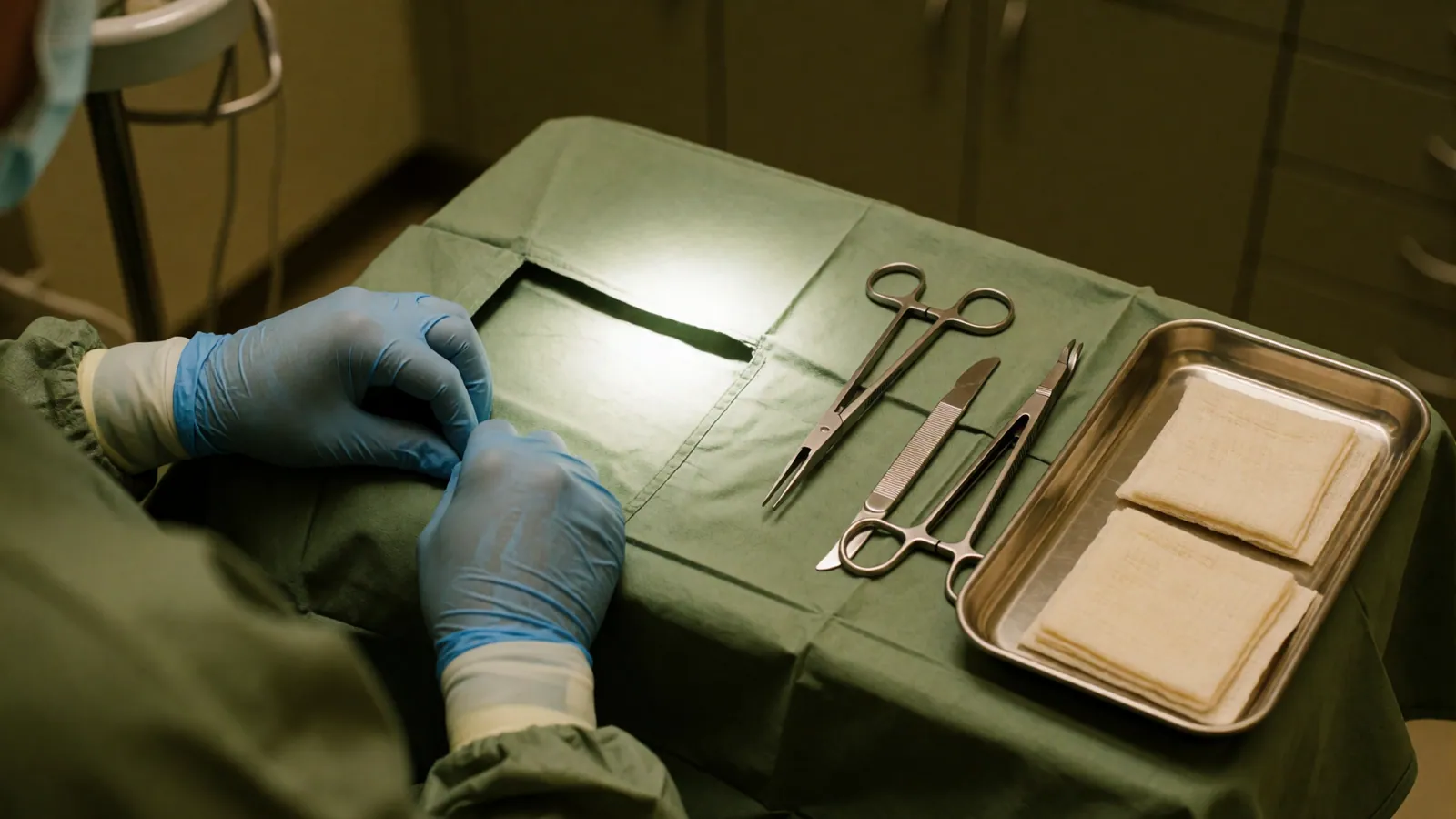
Some things should not wait for the next free routine appointment. Some things should not wait for the next free routine appointment. Telephone the practice and say what is happening, or go to an emergency room, if you have a mole or a mark that has changed shape, colour or size, that has become raised or uneven, or that bleeds, itches or will not heal; a sore anywhere that has not healed in three weeks; a rash that is spreading quickly, especially with fever, blistering, or peeling skin; a painful rash in a band on one side of the body, particularly near an eye; redness that is hot, swollen, tender and spreading, which can be a deep skin infection; a new rash after starting a medicine, especially with mouth or eye involvement; or a child under three months with any widespread rash. If you have been treated here and something is worse rather than better after a procedure, telephone us rather than waiting for the review appointment. We would far rather field a call that turns out to be nothing.
A biopsy removes a small piece of skin so that a pathologist can examine it under a microscope. It is the single most useful procedure in this speciality, because a great many conditions that look alike to the naked eye are entirely distinct under magnification — and because a confident guess and a diagnosis are not the same thing, however experienced the person guessing.
We take biopsies for three reasons. To find out what a rash is when the history and examination have not settled it. To find out whether a growth is a cancer, and if so which. And, occasionally, to prove that something is what we think it is before committing somebody to months of treatment that carries its own risks. In every case, the honest framing is that it is better to know than to watch.
The choice of biopsy technique is not a detail. A rash needs a full-thickness sample, because the pathologist has to see all the layers to see what is happening between them. A raised growth sitting on the surface can be shaved flat. Anything possibly a melanoma must be taken whole with a margin, because the measurement that determines the entire subsequent plan is how deep it goes, and a partial sample can miss the deepest part. Getting this choice right at the outset is most of the value.
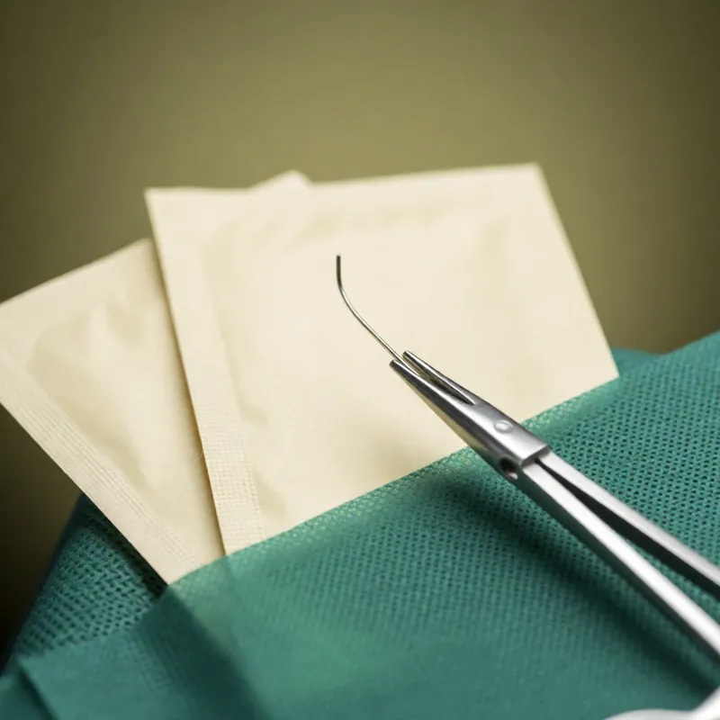
The types, and when each is right
Four techniques, chosen by what question needs answering.
Punch biopsy
A small circular blade, three or four millimetres across, taking a full-thickness core down to the fat. The right choice for a rash, for inflammatory conditions and for the scalp, because it captures every layer. Usually closed with one or two stitches; leaves a pale mark the size of a match head.
Shave biopsy
A blade taking the raised part of a lesion flat to the surface. Quick, no stitches, and appropriate for growths clearly sitting on top of the skin. It leaves a flat, slightly lighter patch. It is the wrong technique for anything that might be a melanoma, and a shaved melanoma frequently leaves the depth unknowable, which matters permanently.
Excisional biopsy
Removing the whole lesion with a margin of normal skin around and beneath it, closed with sutures. This is what anything possibly a melanoma requires, and it is also used where a lesion needs removing anyway.
Incisional biopsy
Taking a wedge from a large lesion or from the edge of a rash where removing all of it would not be sensible. Used for larger areas and for particular inflammatory conditions where the edge is the informative part.
Sometimes two samples, from two places
Particularly for rashes and for scalp biopsies, where a pathologist benefits from comparing sites or from a second sample cut in a different plane. It is not a sign that anything has gone wrong.
What it actually feels like
The part people want described, and the part that is usually skipped.
The anaesthetic is the only part that hurts
A fine needle and a sting for perhaps ten seconds while the anaesthetic goes in. After that the area is numb within a couple of minutes and you should feel pressure and movement but nothing sharp.
Tell us at once if you feel anything sharp
We stop and put more anaesthetic in. This instruction is given to every patient at the start because people endure things unnecessarily rather than interrupt, and there is nothing to be gained by it.
Your heart may race for a minute
The anaesthetic usually contains adrenaline to reduce bleeding, and a brief fluttering or a racing sensation shortly afterwards is expected rather than a reaction.
It takes about fifteen minutes in total
Most of which is consent, cleaning, marking and the anaesthetic taking effect. The sampling itself is quick.
You go home yourself
No sedation, no driver needed. You can go back to work the same day for most sites, avoiding lifting and stretching if the sample is on the trunk.
Say if you have fainted at needles before
It is common, it is not embarrassing, and we do the whole thing with you lying down. Far better said at the start than discovered halfway through.
Healing, results and what happens next
Including the honest bit about scars.
Keep it dry for forty-eight hours, then clean and re-dress
Showering is usually fine after that. No soaking, swimming or hot tubs until any sutures are out.
Sutures out in five to fourteen days
Five to seven on the face, ten to fourteen on the trunk and limbs. Face sutures left too long are what produce the little cross-hatch marks people associate with stitches.
It will leave a mark
A punch leaves a small pale spot; a shave leaves a flat lighter patch; anything sutured leaves a line. On darker skin the site may be darker rather than lighter for many months. On the chest, shoulders and jawline scars are more likely to thicken, and where that applies to you it is said before rather than after.
The result takes about a week
Occasionally longer if the pathologist orders additional stains, which is thoroughness rather than bad news. If you have not heard within ten days, telephone us.
We telephone every result, including the reassuring ones
No news does not mean good news here. Patients who assume it does spend a fortnight assuming the wrong thing, and it is an easy assumption to make.
What is not normal
Increasing pain from day three, spreading redness, heat, swelling, pus or fever. That is how infection presents and it warrants a telephone call rather than waiting for the suture appointment.
Skin Biopsy in western Queens
Taking a small sample of skin so a pathologist can say what is actually happening in it. Fifteen minutes, local anaesthetic, a result in about a week — and we telephone you either way, including when the news is good.
Every appointment here starts the same way: a proper history, an examination in good light, and a named diagnosis said out loud and written down for you to take home. If there is genuine doubt you will be told that, and told what would settle it — usually a biopsy. You leave with a plan that has a strength, a site, a frequency and a date to stop or come back on it, and, where the condition is one that relapses, what to do on the first day of the next flare. If the honest answer is that this is better left alone, or that it belongs somewhere other than here, that is what you get instead, and it costs exactly the same.
This page is not medical advice, and it is not a diagnosis. Nothing on this website is medical advice for you in particular, and it is not a diagnosis. These pages describe conditions in general terms so that you arrive knowing what the questions are — they cannot tell you which condition you have, because that requires somebody to look at your skin, feel it, ask you a hundred questions and frequently take a sample of it. Two conditions on this website can look identical in a photograph and need opposite treatments. If something on your skin is new, changing, painful or not healing, the useful next step is an appointment rather than more reading.
No before-and-after photographs, and no promised outcome. There are no before-and-after photographs on this website and there never will be. Every set you have seen somewhere else was chosen from a much larger pile, and the second picture is usually taken in different light, at a different angle, at a different point in a flare cycle. In dermatology the dishonesty is sharper than in most fields, because nearly every condition here fluctuates on its own — photograph any relapsing skin disease at its worst and again at its best and you have a persuasive pair of images that demonstrates nothing about the treatment in between. What we publish instead is what a treatment typically achieves, in how long, in what proportion of people, and how often it does not work.
The risks, plainly. Every treatment described here has side effects, and some carry real risks. Topical steroids thin skin if they are used at the wrong strength on the wrong site for too long, which is why they are prescribed with a site, a strength and a stopping point rather than handed over indefinitely. Light-based and laser treatment can burn, blister and cause pigment change that lasts months, and that risk is higher on deeper skin tones and on recently tanned skin. Any procedure that breaks the skin can bleed, become infected and leave a scar; on the chest, the shoulders and the jawline, scars are more likely to thicken. Several of the medicines used in this speciality require blood tests, and some cannot be taken during pregnancy. We tell you your own risk rather than the average one, and where the honest answer is that a condition is better left alone, that is the advice you will get.
Nothing is diagnosed from a photograph. We do not diagnose skin from photographs, and we do not answer "what is this?" by email, by message or through a form. This is not caution for its own sake. A photograph has no history attached to it, cannot be felt, cannot be stretched or pressed to see whether the colour blanches, cannot be examined under a dermatoscope, and misreports colour badly — which matters enormously on skin that is not pale. The examination that actually distinguishes a harmless growth from an early cancer involves magnification, cross-polarised light and, when there is any doubt at all, a biopsy. An application that offers to tell you whether a mole is dangerous from a picture taken on a telephone is selling reassurance, and reassurance is the one product in this field that is dangerous when it is wrong. Bring the mark to a room where somebody can look at it.
Questions
Skin Biopsy — the questions we are asked most
Will it hurt?
The anaesthetic stings for about ten seconds. After that you feel pressure and movement but nothing sharp. If you do feel anything sharp, say so immediately and we put more in — nobody should be enduring it quietly, and patients frequently do.
Will it leave a scar?
Yes, some mark. A punch biopsy usually heals to a pale spot the size of a match head, a shave to a flat lighter patch, and anything sutured to a line. On darker skin the site may be darker rather than lighter for months. Where a site is prone to thickened scarring you will be told beforehand and we will discuss whether to sample somewhere else.
Can I take my blood thinner beforehand?
Yes, unless the doctor who prescribed it has told you otherwise. The bleeding risk from a small skin sample is manageable; stopping an anticoagulant without instruction is not. Tell us what you take, including aspirin and supplements such as fish oil.
How long until I get the result?
About a week, sometimes longer if extra stains are needed. We telephone every result, good or bad, and you will be told before you leave how and when that call will come. If ten days pass without a call, telephone us — do not assume.
Why do I need a biopsy when you already think you know what it is?
Because thinking and knowing are different, and the difference matters when the next step is months of treatment with its own risks, or when the alternative diagnosis is a cancer. A biopsy that confirms what we expected is a good outcome rather than a wasted procedure.
Could taking a sample make a cancer spread?
No. This is a common and understandable worry and it is not what happens. Sampling does not cause a cancer to spread, and what genuinely does worsen outcomes is delay in diagnosis.
Related
Often seen alongside this
Procedures & Testing
Cryotherapy
Liquid nitrogen, applied in short bursts, to freeze off sun-damage patches, warts and several harmless growths. Quick and effective. It stings for twenty seconds and it usually leaves a pale mark — which on deeper skin tones can be permanent.
Read more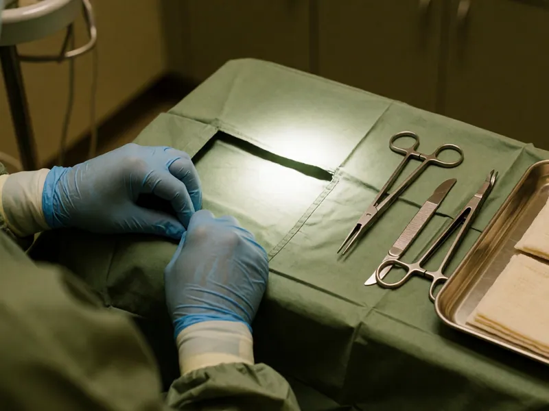
Procedures & Testing
Cyst & Lipoma Removal
Removing a cyst or a fatty lump under local anaesthetic. Both are harmless; both are worth removing when they are growing, catching or repeatedly inflamed. The timing matters — an inflamed cyst is settled first and removed later.
Read more
Procedures & Testing
Patch Testing
Panels of standardised allergens taped to the back for two days and read twice across a week. Unglamorous, slow, and the only reliable way to find out what you are genuinely allergic to. It ends more long-running dermatitis than anything else in this building.
Read moreA named diagnosis and a written plan you leave holding
Book an appointment
Pick a physician and a time from the calendar — it shows only the hours each of them genuinely has free. A new patient appointment is thirty minutes, a full skin examination forty-five, and you leave with a named diagnosis written down. If something on your skin has changed, telephone instead and say that word: it is triaged differently.